İçeriğe atla

Dosya:3D-fluorescence imaging for high throughput analysis of microbial eukaryotes.jpg

Sayfa içeriği diğer dillerde desteklenmemektedir.
Vikipedi, özgür ansiklopedi

Tam çözünürlük((5.098 × 3.285 piksel, dosya boyutu: 1,39 MB, MIME tipi: image/jpeg))


Özet

Açıklama
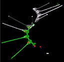
English: Environmental high-content fluorescence microscopy (e-HCFM): automated, 3D, and multichannel imaging for aquatic micro-eukaryotes.
(a) e-HCFM workflow applied to Tara Oceans samples: (1) 72 nano-plankton (size range 5–20 μm) samples collected during the Tara Oceans expedition (Pesant et al., 2015) were fixed in paraformaldehyde-glutaraldehyde buffer onboard and kept at 4°C for up to several years; (2) Samples were mounted in optical multiwell plates. Then, a 4-steps preparation allowed to stain all eukaryotic cells; (3) A commercial confocal laser scanning microscope was used to automatically image samples (40X NA1.1 water lens; 5 channels) generating 2.5 Tb of raw data (acquisition details in Figure 1—source data 1); (4) In total, 336,655 objects were processed for individual extraction of 480 descriptors (3D biovolumes, intensity distribution, shape descriptors and texture features, details in Figure 1—source data 2), and the reconstruction of various images for visual inspection (c); (5) A training set based on 18,103 manually curated images (5.4% of the dataset) classified into 155 categories, was used for automated recognition (Random Forest). (b) Examples of e-HCFM 3D-images and movies from a wide phylogenetic diversity of planktonic eukaryotes (see also Figure 1—figure supplements 1 and 2). Left panel: a chain of diatoms (Asterionellopsis sp., Heterokonta) (Figure 1—video 1); right panel, top left to bottom right: radiolarian (Rhizaria), ciliate (Alveolata), amoeba (Amoebozoa), choanoflagellate (Opisthokonta), dinoflagellate (Alveolata), coccolithophore (Haptophyta). Key cellular features are labelled with various dyes: DNA/nuclei (blue, Hoechst33342); (intra)cellular membranes (green, DiOC6(3)); cell covers and extensions (cyan, PLL-AF546, a home-made conjugation between α-poly-L-lysine (PLL) and Alexa Fluor 546 (AF546)); chloroplasts (red, chlorophyll autofluorescence). Scale bar 5 µm. (c) The confocal microscope is automated for acquiring multicolor Z-stacks over a mosaic of positions for each sample.
Tarih
Kaynak

[1]

doi:10.7554/eLife.26066.003
Yazar Sebastien Colin, Luis Pedro Coelho, Shinichi Sunagawa, Chris Bowler, Eric Karsenti, Peer Bork, Rainer Pepperkok, Colomban de Vargas
İzin
(Bu dosyanın tekrar kullanımı)

This article is distributed under the terms of the Creative Commons Attribution License, which permits unrestricted use and redistribution provided that the original author and source are credited.

Diğer sürümler

Lisanslama

w:tr:Creative Commons
atıf
Bu dosya Creative Commons Atıf 4.0 Uluslararası lisansı altında lisanslanmıştır.
Şu seçeneklerde özgürsünüz:
  • paylaşım – eser paylaşımı, dağıtımı ve iletimi
  • içeriği değiştirip uyarlama – eser adaptasyonu
Aşağıdaki koşullar geçerli olacaktır:
  • atıf – Esere yazar veya lisans sahibi tarafından belirtilen (ancak sizi ya da eseri kullanımınızı desteklediklerini ileri sürmeyecek bir) şekilde atıfta bulunmalısınız.

Altyazılar

Bu dosyanın temsil ettiği şeyin tek satırlık açıklamasını ekleyin.
3D-fluorescence imaging for high throughput analysis of microbial eukaryotes

Bu dosyada gösterilen öğeler

betimlenen

31 Ekim 2017

image/jpeg

6d5081452264c5af9eb495f60026672fcbfcfc07

1.452.947 Bayt

3.285 piksel

5.098 piksel

Dosya geçmişi

Dosyanın herhangi bir zamandaki hâli için ilgili tarih/saat kısmına tıklayın.

Tarih/SaatKüçük resimBoyutlarKullanıcıYorum
güncel22.11, 19 Ekim 202022.11, 19 Ekim 2020 tarihindeki sürümün küçültülmüş hâli5.098 × 3.285 (1,39 MB)RemitamineHigher resolution version
07.57, 5 Ekim 202007.57, 5 Ekim 2020 tarihindeki sürümün küçültülmüş hâli1.500 × 966 (256 KB)EpipelagicUploaded a work by Sebastien Colin, Luis Pedro Coelho, Shinichi Sunagawa, Chris Bowler, Eric Karsenti, Peer Bork, Rainer Pepperkok, Colomban de Vargas from [https://elifesciences.org/articles/26066] {{doi|https://doi.org/10.7554/eLife.26066.003}} with UploadWizard

Bu görüntü dosyasına bağlanan sayfa yok.

Küresel dosya kullanımı

Aşağıdaki diğer vikiler bu dosyayı kullanır: